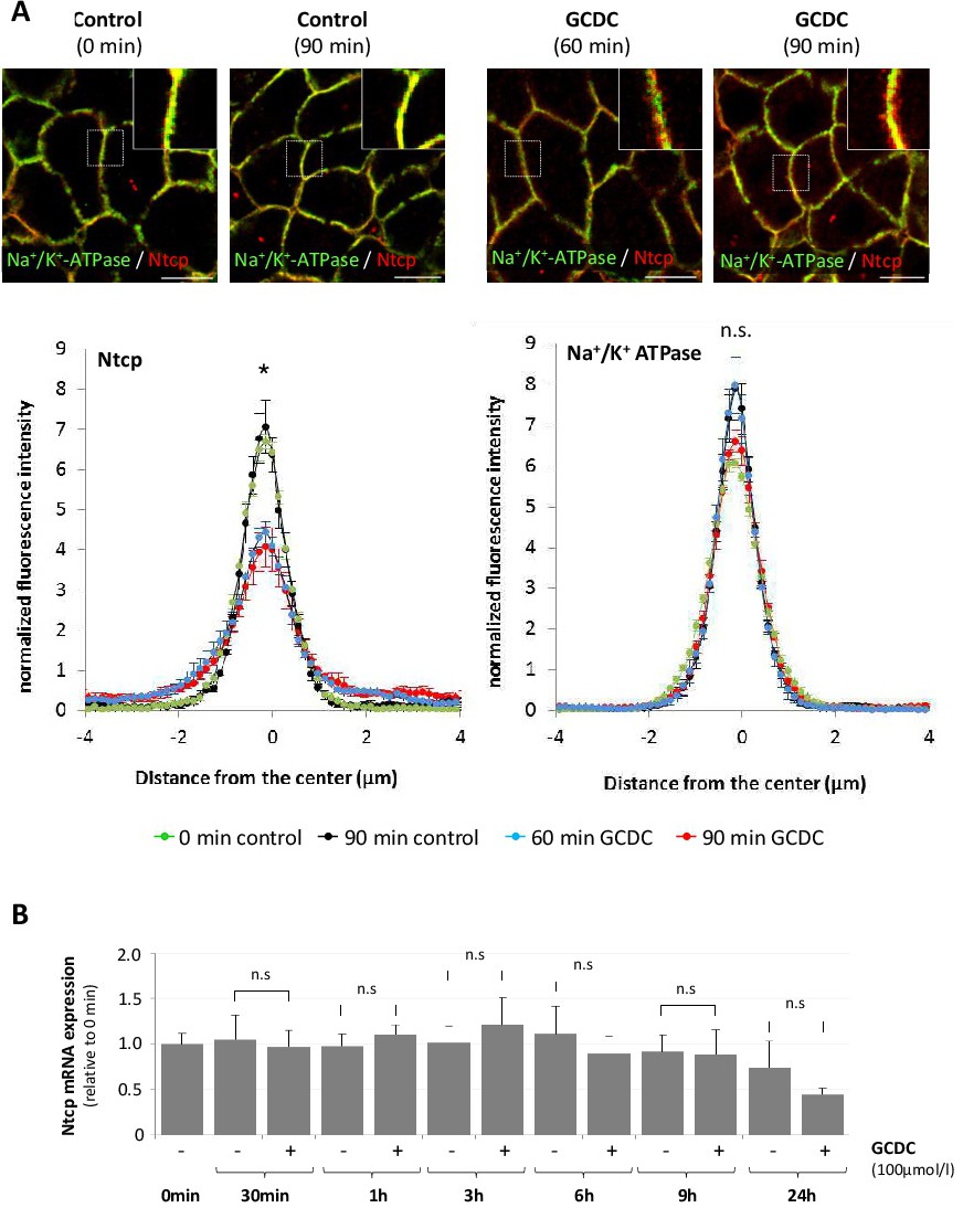
Fig. 1. Effects of GCDC on Ntcp protein localization in rat liver and Ntcp mRNA levels in cultured hepatocytes. (A) Livers were perfused with Krebs-Henseleit buffer with or without GCDC (20 µmol/l) for up to 90 min. Cryosections of livers were immunostained for Ntcp and Na+/K+-ATPase. Fluorescence pictures were acquired by confocal laserscanning microscopy and fluorescence distribution was measured over a line (8 µm) perpendicular to the plasma membrane of adjacent hepatocytes as described in the methods section. Magnifications of the boxed regions are shown in the upper right corner of each picture. Data represent arithmetric means ± SEM of 10 measurements in each of at least 3 individual experiments for each condition. (B) Primary rat hepatocytes were cultured for 24 h and thereafter exposed to GCDC (100 µmol/l) for up to 24 h. RNA was extracted and Ntcp mRNA expression levels were analyzed by realtime-PCR. Ntcp mRNA expression levels are given relative to the untreated control (0 min). Data represent arithmetic means ± SEM of 4 independent experiments. * statistically significantly different compared to control. n.s.: not statistically significantly different.